 by
by
× nearby
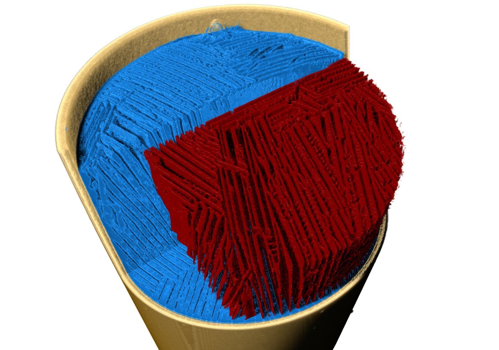
The 3D-rendered tomogram shows a cross-section through a solidified sample where two phases have separated: the pure ice crystal phase in green and the sugar phase in red. The lamellar structure formed by snowflake-like ice crystals is clearly visible. Credit: HZB/PSI
Freeze casting processes can be used to produce highly porous and hierarchically structured materials. They are suitable for a variety of applications, such as electrodes for batteries, assistive devices or in biomedicine.
Now the group led by Prof. Ulrike GK Wegst, Northeastern University, Boston, MA, US and Dr. Francisco García Moreno from the Helmholtz-Zentrum Berlin have now used a new X-ray tomoscopy technique at the Swiss Light Source of Paul. Scherrer Institute to observe in real time and in high resolution how the building process was done during the ice age. A sugar solution served as a model system.
Freeze casting requires several steps: First, the material is melted or suspended in a solvent and then frozen in a mold with the cooling rate set at the bottom (solidification of the guide). After freezing, the solid solvent phase is removed by sublimation. What remains are pre-dissolved molecules and suspended particles, which form cell walls of a complex, highly porous effect.
Cast iron materials can be used in many applications: for example, because of their large internal surfaces as battery electrodes or catalysts or because of their relative porosity in biomedical applications for example as scaffolds for peripheral nerve repair. However, exactly how the ice exhibits a complex structure during freezing, and how the desired honey-like porosity and cell walls with their different surface properties, has been poorly understood so far.
Dr. Francisco García Moreno and his team at the Helmholtz-Zentrum Berlin have developed a method to observe these highly dynamic processes in detail. “Using X-ray tomography, we can image the formation of structures in the area with a high resolution of space and time and observe transitory and transitional structures,” explains the physicist.
Using an ultrafast turntable, sharp X-rays, a very fast detector and software for rapid analysis of X-ray data, the HZB team, together with colleagues from the Swiss Light Source of the Paul Scherrer Institute, learned how to throw ice into the model. system and show high performance of the method.
× nearby
The bottom of the mold is cooled on a turntable with liquid nitrogen and simultaneously analyzed with X-rays. Ice crystals grow in the “cold finger” and grow in the direction of temperature. During solidification, dissolved or suspended material is concentrated in the interstices. Credit: HZB/PSI
“In this study, we created a new measuring cell and sensors to accurately record the temperature gradient,” said Dr. Paul Kamm (HZB), lead author of the study. A 3D tomogram with a spatial resolution of 6 µm per second was generated. The entire freezing process was recorded for over 270 seconds.
Prof. Ulrike GK Wegst from Northeastern University, US, proposed the aqueous sugar solution as a polymeric model system, since this process can be measured computationally, and because water solutions still dominate the freezing process. “We are now able to observe experimentally for the first time the dynamics of lead ice crystals growing from the liquid phase,” said Wegst.
“In doing so, the images document the instability during crystal growth, how these form sugar phases and how characteristic, organic-looking structures are formed in the cell walls reminiscent of jellyfish and tentacles.” It is also interesting to note that some of these buildings may disappear again.
This paper is published in a journal High Performance Materials.
More information:
Paul H. Kamm et al, X-Ray Tomoscopy Reveals the Dynamics of Ice Templating, High Performance Materials (2023). DOI: 10.1002/adfm.202304738
Journal information:
High Performance Materials
#formation #structures #ice #casting #photographed #real #time